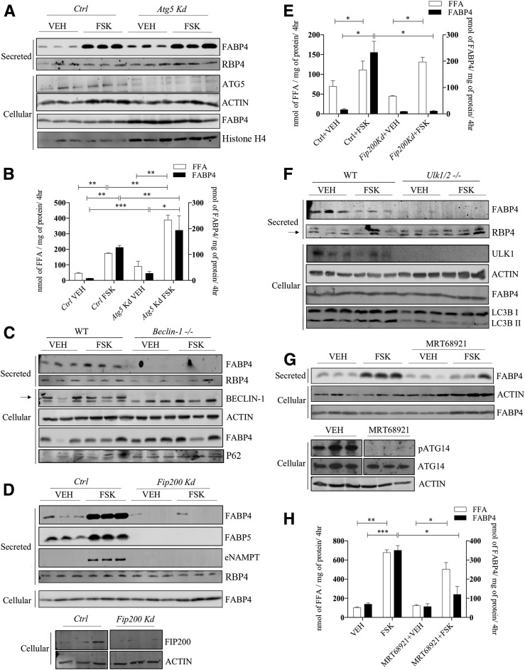
Figure 3.
FABP4 secretion in response to molecular regulators of early autophagic proteins. A: Lentiviral-infected control (Ctrl) or Atg5-silenced 3T3-L1 adipocytes were treated with VEH or 20 μmol/L FSK for 4 h, and the secretion of FABP4 and RBP4, as well as the cellular levels of ATG5, β-actin, FABP4, and histone H4, was evaluated immunochemically. B: Quantitative analysis of the levels of secreted FFA and FABP4 from A. C: MEFs from a wild-type C57BL/6J or beclin-1 null mouse were differentiated as described in research design and methods, and secreted FABP4 and RBP4 and cellular levels of beclin-1, β-actin, FABP4, and P62 were determined immunochemically. D (top): Lentiviral-infected control or Fip200-silenced 3T3-L1 adipocytes were treated with VEH or 20 μmol/L FSK for 4 h, and the secretion of FABP4, FABP5, eNAMPT, and RBP4 as well as cellular FABP4 was determined immunochemically. D (bottom): Cellular levels of FIP200 and β-actin in control or Fip200-silenced 3T3-L1 adipocytes. E: Quantitative analysis of FABP4 (from D) and FFAs secreted by control or Fip200-silenced 3T3-L1 adipocytes. F: MEFs from wild-type C57BL/6J or ULK1/2 null mice were differentiated and the expression of secreted FABP4 and RBP4 and cellular levels of ULK1/2, β-actin, FABP4, and LC3B were determined immunochemically. The arrow indicates specific RBP4 band. G: Differentiated 3T3-L1 adipocytes were treated with or without 2 μmol/L of the ULK1/2 inhibitor MRT68921 for 2 h followed by addition of either VEH or 20 μmol/L FSK for 4 h. The levels of secreted FABP4 and cellular β-actin and FABP4 were evaluated immunochemically. Additionally, the cellular levels of phosphorylated ATG14 (pATG14), ATG14, and β-actin were evaluated immunochemically. H: Quantitation of FABP4 and FFA secreted by 3T3-L1 adipocytes in response to VEH or 20 μmol/L FSK stimulation in the presence and absence of the ULK1/2 inhibitor MRT68921. *P < 0.05; **P < 0.01; ***P < 0.001. hr, hours; kd, knockdown.

