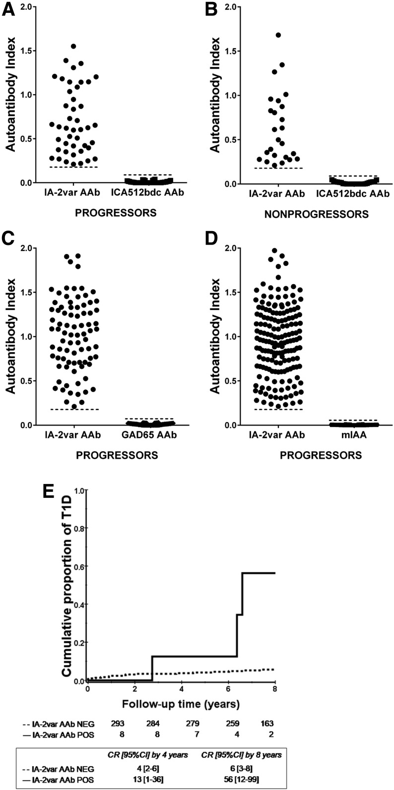
Figure 4.
IA-2var AAb can be detected in relatives who are negative for traditional islet AAb at screening (ICA512bdc, GAD65, and mIAA). A: 7.8% (44 of 566) of the relatives who developed T1D (progressors) were negative for ICA512bdc but negative for IA-2var AAb. B: 2.2% (25 out of 1,120) of ICA512bdc AAb–negative nonprogressors were IA-2var AAb positive. C and D: 14.2% (81 of 566) of progressors were IA2var AAb positive and GAD65 AAb negative (C), and 29.7% (168 of 566) of the progressors were IA-2var AAb positive but mIAA negative (D). E: In seronegative relatives at screening (negative for ICA512bdc, GAD65 AAb, IAA), the presence of IA-2var AAb (solid line) still conferred a higher risk of progression to T1D compared with those negative for IA-2var AAb (dashed line) (P = 0.001). CR, cumulative risk; NEG, negative; POS, positive.

