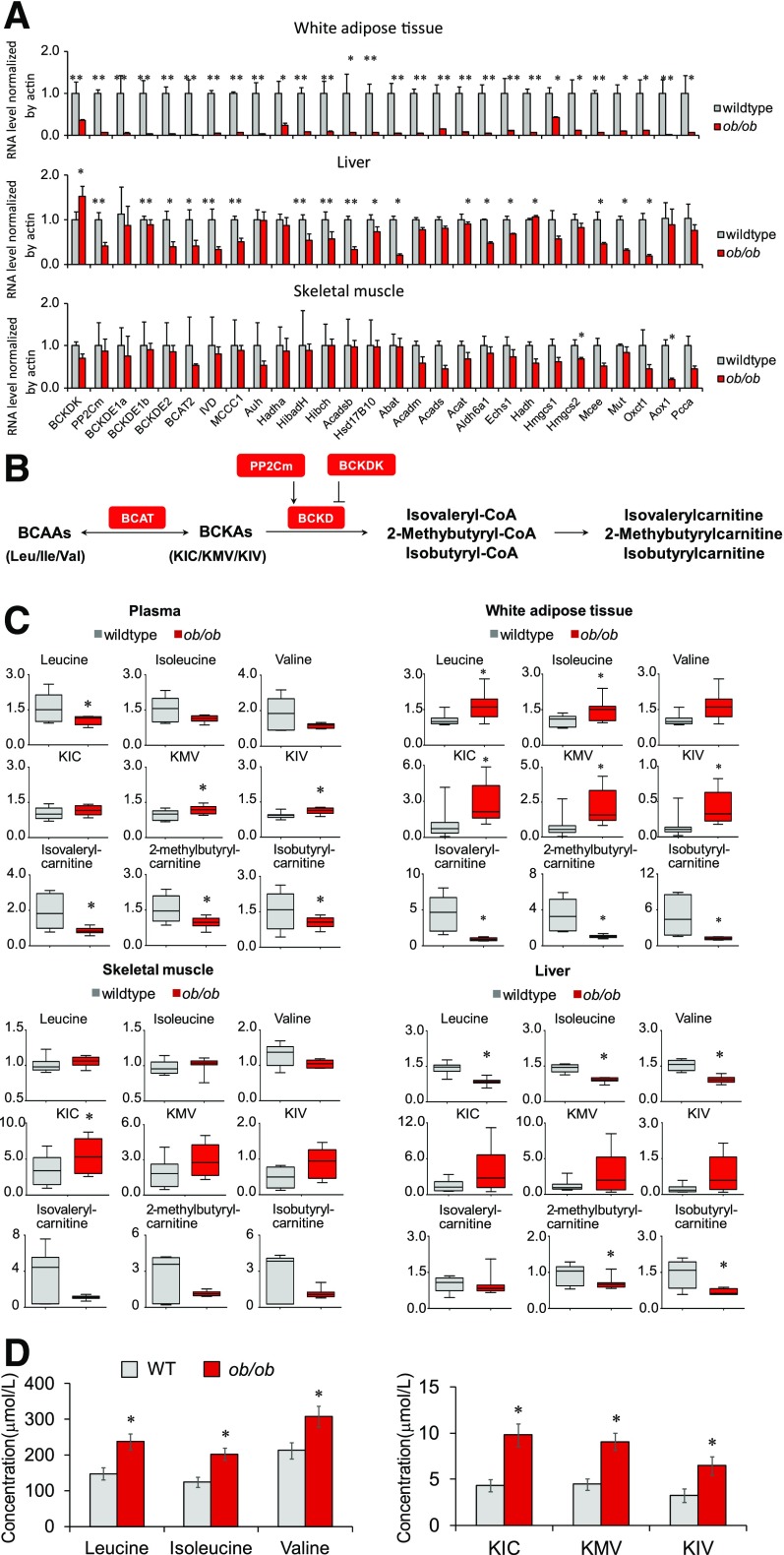
Figure 3.
Systematic downregulation of the BCAA catabolic pathway leads to a BCAA catabolic defect in ob/ob mice. A: Quantitative PCR results of BCAA catabolic genes in white adipose tissue, skeletal muscle, and liver in lean wild-type (WT) mice (n = 4) and ob/ob mice (n = 4) deprived of food for 6 h. B: Illustration of the partial BCAA catabolic process with enzymes, intermediates, and derivatives. C: Relative levels of BCAAs and their metabolites in plasma and tissues of lean WT mice (n = 6–8) and ob/ob mice (n = 8). Male mice, age 14 weeks, were deprived of food for 6 h before being sacrificed. D: Serum concentrations of BCAAs and BCKAs in lean WT mice (n = 8) and ob/ob mice (n = 10). Male mice, age 8 weeks, were deprived of food from 8:00 a.m. to 5:00 p.m. and then supplied with chow diet for 1 hour. Blood (100 μL) was collected at 6:00 p.m. from the orbital sinus by using capillary tubes for serum collection. *P < 0.05 and **P < 0.01 vs. WT mice. ΚΙC, α-ketoisocaproic acid; KIV, α-ketoisovaleric acid; KMV, α-keto-β-methylvaleric acid.

