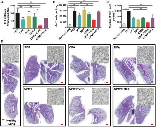
Figure 3.

Combination therapy using CPMV and CPA reduces lung metastasis. A) Normalized absorbance of crystal violet in different treatment groups (n = 4). (Lungs were harvested 8 days after the first treatment.) B) The number of Gr‐1+ granulocytes in the lung (n = 4, harvested 8 days after the first treatment) determined by flow cytometry. C) Abundance of dividing cells (Ki67 positive) in the lung sections (n = 2). D) Representative lung sections stained with H&E (Scale bar (red) = 500 µm). The inserted images are the representative immunochemical stain of Ki67 antibody (Scale bar = 100 µm). The data represent means and standard deviations, with statistical analysis by one‐way ANOVA (ns = not significant, **p < 0.01).
