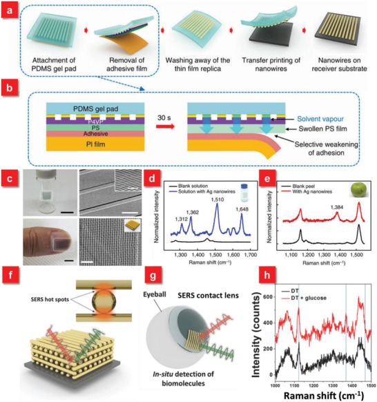
Figure 7.

a) Procedure for solvent‐assisted nanotransfer printing. b) The mechanism of transferring the nanostructures and the polymer replica to the surface of PDMS gel pad by removal of the adhesive film. c) S‐nTP of metallic nanowires transferred onto arbitrary surfaces. d) Raman signal spectra obtained from a vial glass containing 10−5 m R6G solution. e) Raman signal obtained from the surface of an apple coated with Thiram (tetramethylthiuram disulfide) with an areal density of 1 µg cm−2. Raman signal peak at 1384 cm−1 corresponds to that of Thiram. Reproduced with permission.42 Copyright 2014, Nature Publishing Group. f) A schematic diagram of 3D cross‐point plasmonic nanostructures to enhance SERS performance. g) A schematic illustration of SERS contact lens via transfer printing for in situ detection. h) Comparison of SERS spectra before and after dropping glucose solution, showing the successful detection of glucose. Reproduced with permission.45 Copyright 2016, John Wiley & Sons.
