Figure 1.
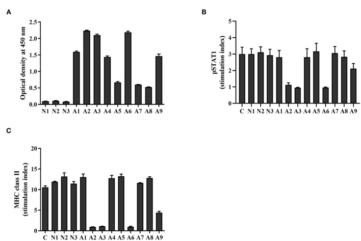
Level and neutralizing ability of anti-IFN-γ autoAbs in AOID sera. (A) Anti-IFN-γ autoAbs in patient sera [cohort; patients with opportunistic infections (n = 9) and healthy controls (n = 3)] were measured by indirect ELISA. The neutralizing capacity of anti-IFN-γ autoAbs was assessed with a cell-based assay. THP-1 cells were treated with IFN-γ in the presence or absence of patient serum. (B) The intracellular pSTAT1 and (C) the surface MHC-II expression was evaluated by flow cytometry. The stimulation index was the ratio of the fluorescence intensity of those proteins in stimulated cells divided by that in unstimulated cells. Experiments were performed in triplicate. Error bars represent the mean ± SEM.

