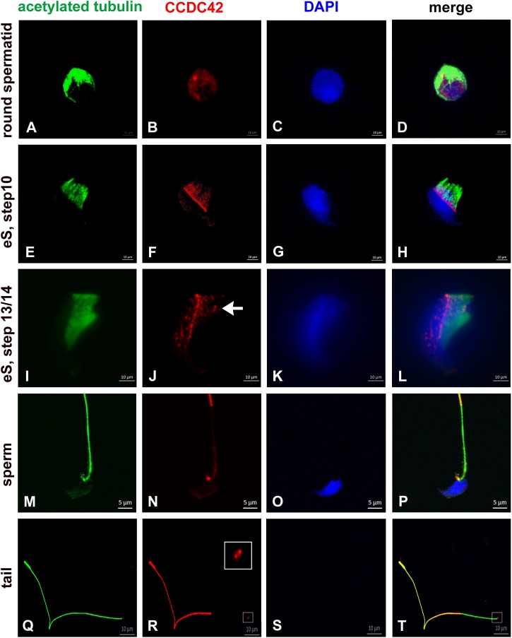
FIGURE 1.
CCDC42 localizes in the manchette, the perinuclear ring and the connecting piece during spermiogenesis. Suspension preparations of adult mouse testis were incubated with antibodies against acetylated tubulin (green) and CCDC42 (red). Weak staining for CCDC42 was observed in the cytoplasm in round spermatids (A–D). In elongating spermatids (eS) CCDC42 decorated the manchette and more strongly the perinuclear ring (E–L). Additionally, the HTCA or connecting piece harbored CCDC42 as well (I–L, arrow in J). CCDC42 also located to the connecting piece in sperm (M–P) and to the tail with highest expression in the principal piece (M–T). In detached tails, the anterior region, which corresponds to the attachment site to the nucleus, stained for CCDC42 (Q–T; framed in R and S and inset in R showing the enlarged region). Secondary antibodies used are anti-mouse IgG-Dylight488 and anti-rabbit IgG-MFP590 (A–H,M–P) or anti-rabbit IgG-Dylight488 and anti-mouse IgG-Alexa Fluor R 555 (I–L,Q–T). Nuclear staining with DAPI (blue). Bars are of 10 μm except for M–P in which 5 μm scales are used.

