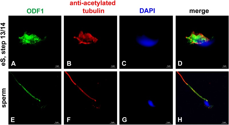
FIGURE 4.
ODF1 locates to the manchette in elongating spermatids and to the sperm tail. Incubation of mouse testicular cells with antibodies against ODF1 (green) and acetylated tubulin (red). ODF1 decorates the manchette in elongating spermatids (A–D) and the sperm tail (E–H). Nuclear counterstain with DAPI (blue). Bars: 2 μm (A–D), 5 μm (E–H).

