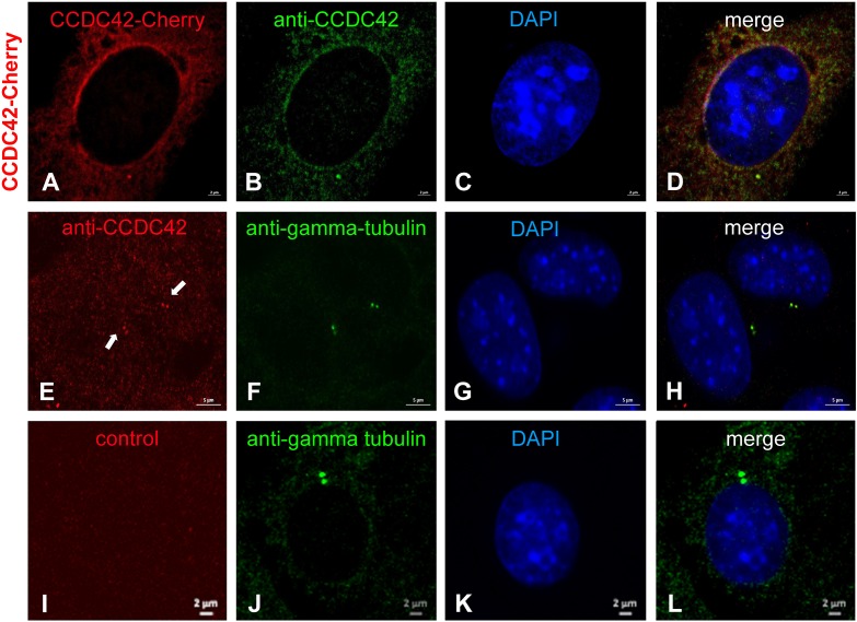
FIGURE 9.
CCDC42 is a centrosomal component in NIH3T3 cells. (A–D) NIH3T3 cells were transfected with Ccdc42-Cherry expression plasmid (as indicated on the left side by CCDC42-Cherry) and CCDC42 detected by its fluorescent tag (CCDC42-Cherry, red) and immuno-decoration (anti-CCDC42, green) to validate the antibody. (E–H) Endogenous expression of CCDC42 in NIH3T3 cells was detected immunocytologically (anti-CCDC42, red) and co-located with the centrosomal marker protein γ-tubulin (anti-gamma-tubulin, green). (I–L) Omitting anti-CCDC42 antibody incubation showed no red decoration of the centrosome, which was otherwise detected by anti-γ-tubulin staining (anti-gamma-tubulin, green), demonstrating anti-CCDC42 antibody specificity. Nuclear counterstain with DAPI (blue). Bars are of 2 μm (A–D,I–L) or of 5 μm (E–H).

