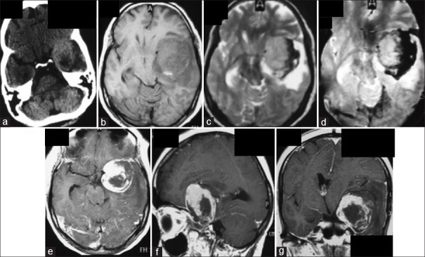
Figure 1.
Computed tomography scan of the head performed after the acute episode of a headache showing an ovoid left medial temporal mass lesion. The hyperdensity is not uniform, and there is a pocket of hypodense area posteriorly (a). The lesion on magnetic resonance imaging appears bigger and is hypointense on T1 except for a small hyperdensity posteriorly, whereas it is isointense on T2 image and capped by a hypointense area toward the cerebral convexity, the latter showing blooming on susceptibility-weighted image (b-d). The lesion shows strong but inhomogeneous enhancement (e-g). The center and the posterolateral periphery of the lesion does not enhance at all. Moreover, the nonenhancing portions are capped by another layer of nonenhancing tissue, possibly blood clot (e-g)

