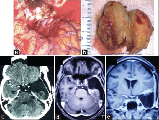
Figure 2.

Postexcision surgical cavity with an internal carotid artery with its bifurcation, optic nerve, and oculomotor nerves visualized at the free edge of the tentorium (a). The specimen showing hemorrhage peripherally with an otherwise fleshy, nonnecrotic interior (b). Postoperative computed tomography (done within 24 h of surgery) as well magnetic resonance imaging (done after two months) showing evidence of gross total tumor excision (c-e)
