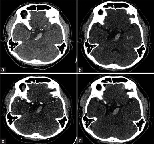
Figure 2.

Computed tomography imaging of a 51-year-old male with underlying hypertension and hyperlipidemia who presented with dysarthria and right facial palsy. (a) Initial computed tomography scan showed hyperdensity and dolichoectasia of the basilar artery. (b-d) Hounsfield unit value was approximately 75, 71, and 70 on noncontrast computed tomography, computed tomography angiography, and contrast-enhanced computed tomography, respectively. The patient was sent for mechanical thrombectomy, the histopathologic results showed mixed thrombus, and the 90-day modified Rankin scale was 4
