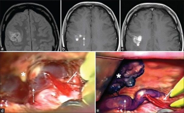
Figure 1.
Magnetic resonance imaging. (a) Axial DP sequence. (b and c) Axial SE T1-weighted after contrast administration. (d) Operative photograph showing the cavernous angioma (asterisk) fine arterial radicles (continuous arrow) entering the main venous collector of the developmental venous anomalies (dotted arrow). (e) The artistic drawing representing the cavernous angioma (asterisk) and the arterialization (continuous arrow) of the main venous channel of the developmental venous anomalies (dotted arrow)

