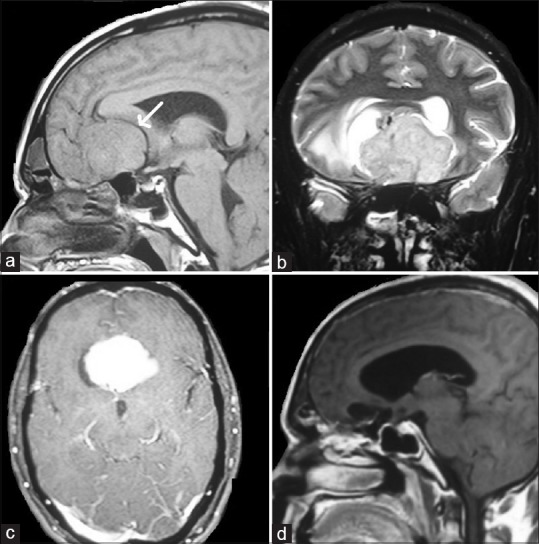
Figure 4.

(a) T1-weighted sagittal section magnetic resonance imaging brain with isointense lesion in the planum sphenoidale, white arrow indicates anterior cerebral artery being pushed upward and posteriorly by the lesion. (b) T2-weighted coronal section magnetic resonance imaging brain with heterointense lesion the planum sphenoidale. (c) T1-weighted postcontrast axial section magnetic resonance imaging brain with contrast enhancing lesion in the planum sphenoidale. (d) T1-weighted postcontrast sagittal section magnetic resonance imaging brain postoperative image with complete excision of the lesion
