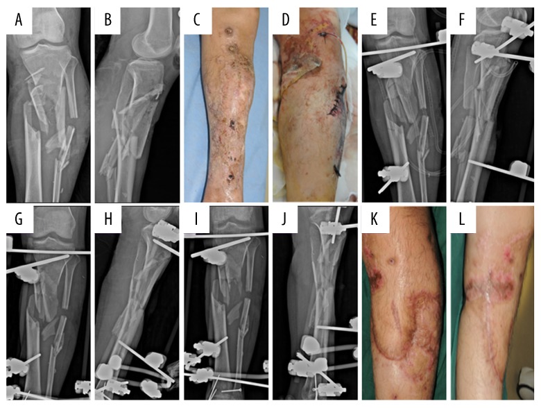
Figure 2.
A male patient with open comminuted fracture of the left tibiofibular and upper leg degloving injury. (A, B) Radiography showed comminuted fracture of the upper segment of the left tibiofibula with obvious displacement. (C, D) Soft tissue condition indicated the anterior and lateral skins of the upper left leg were torn and sutured and extensively leathered. (E, F) Postoperative radiography after emergency second-stage clearance showed that the position of the external fixator was acceptable. (G–L) Radiographic examinations showed poor alignment, and no callus formation at 1 and 3 months after surgery, as well as extensive scarring on the medial part of the upper leg.

