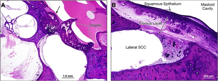
Figure 2.

Right ear, hematoxylin and eosin (H&E) stain. (A) Scaled section showing fixation of the thickened stapes footplate with an anterior focus of otosclerosis (denoted by the arrow). Moderate to severe loss of spiral ganglion neurons is evident. (B) Section at twice the previous magnification demonstrating the lateral SCC fenestration with a thick overlying fibrous layer and squamous epithelium. The fenestration was patent, as bony regrowth had not occurred to close the window. SCC = semicircular canal.
