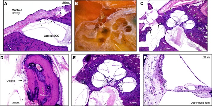
Figure 3.

Left ear, hematoxylin and eosin (H&E) stain. (A) Similar to the right ear, there is a patent lateral SCC fenestration between mastoid cavity (left) and lateral SCC (right). A thick fibrous layer covered by squamous epithelium separates the two spaces. Some bone regrowth is evident (arrows). (B) Specimen, during sectioning and prior to staining, demonstrating previous stapedectomy with a wire loop prosthesis attached to the malleus and extending to the oval window fenestration. (C) Section showing otosclerosis (indicated by arrows) anterior and posterior to the oval window, surrounding the cochlea and anterior to the internal auditory canal. The stapes footplate is absent, suggesting the patient likely had a total stapedectomy. (D) Section of the malleus with osteitis from the wire loop prosthesis. (E) Cochlea showing degeneration of spiral ganglion neurons. (F) Section of the upper basal turn of the cochlea showing some outer hair cell loss and degeneration of the stria vascularis. SCC = semicircular canal.
