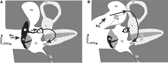
Figure 4.

Two‐dimensional schematics of three‐dimensional views of the right middle ear from a superior view. (A) In the normal middle ear, ear‐canal sound pressure (PEC) produces ossicular motion that drives the cochlear lymphs (shaded with diagonal lines). The motion of the nearly incompressible lymphs and the cochlear partition (CP) depends on the low‐impedance of the round window (RW) that moves in the opposite direction from the stapes (S). (B) In an otosclerotic ear with fixed stapes (X), after a canal‐wall down exposure of the mastoid antrum (labeled An), removal of the incus and fenestration of the lateral canal (LC) covered by a flap of ear canal skin, sound pressure directly moves the lymphs behind the skin‐covered fenestra. This motion is again balanced by an opposite motion of the RW. Other labeled anatomical structures include: ET = eustachian tube; I = incus; M = malleus; ST = scala tympani; TC = tympanic cavity; TM = tympanic membrane; V = vestibule of the inner ear.
