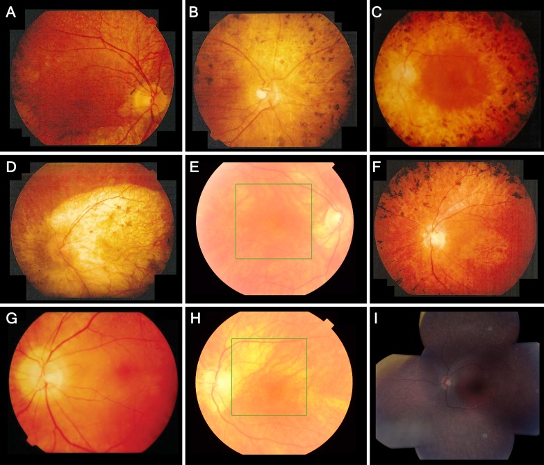
Figure 2.
Fundus photographs showing retinal features and intrafamilial variability in patients with LRAT-associated retinal degeneration. Reported ages are at the time of fundus photography. (A) Patient II, aged 39, showing pigmentary changes in the posterior pole, a macular sheen, and yellow-white dots, associated with retinitis punctata albescens, in the midperiphery. The periphery, not shown here, showed extensive bone-spicule hyperpigmentation. (B) Patient IV, aged 49, showing a pale optic disc, peripapillary atrophy, vascular attenuation, mottled retinal pigmentary epithelium (RPE) changes and clumping in the posterior pole, and atrophy in the nasal midperiphery. In the inferior midperiphery, not shown here, paving stone–like atrophic zones were seen with fundoscopy. (C) Patient VI, aged 42, showing retinal atrophy, and widespread bone spicule–like and coarse hyperpigmentation extending into the posterior pole. The central macula is relatively spared, with mild RPE alterations. (D) Patient I, aged 50, showed profound chorioretinal atrophy of the posterior pole and along the vascular arcade. The periphery, not shown here, showed mild RPE mottling, several paving stone–like degenerations, and little bone spicule–like hyperpigmentation. (E) Patient XII, aged 56, showing peripapillary atrophy and areas of retinal thinning along the superior vascular arcade, with no RPE changes in the central retina. In the inferonasal midperiphery, not shown here, a few round and bone spicule–like pigmentations were clustered. (F) Patient V, aged 47, showed atrophic RPE alterations and a sheen in the central macula, atrophy around the optic disc and along the vascular arcade, and extensive bone spicule–like pigmentation in the midperipheral retina. (G, H) Patient XI, showing the disease progression between the ages of 43 (G) and 67 (H). At the age of 43, this patient showed peripapillary atrophy, yellow–white dots below the superior vascular arcade, and no RPE changes of the central macula. At the age of 67, the white dots were no longer visible, and the posterior pole showed zones of atrophic RPE. (I) Composite fundus photography of the left eye of patient XIII at the age of 15, showing peripapillary atrophy, a normal macula, limited intraretinal white dots in the superior macula that appear less brightly yellow and less sharply circumscribed than the white–yellow dots seen in patients from the genetic isolate, and fleck-like outer retinal atrophy in the (mid-)periphery.

