
Figure 3.
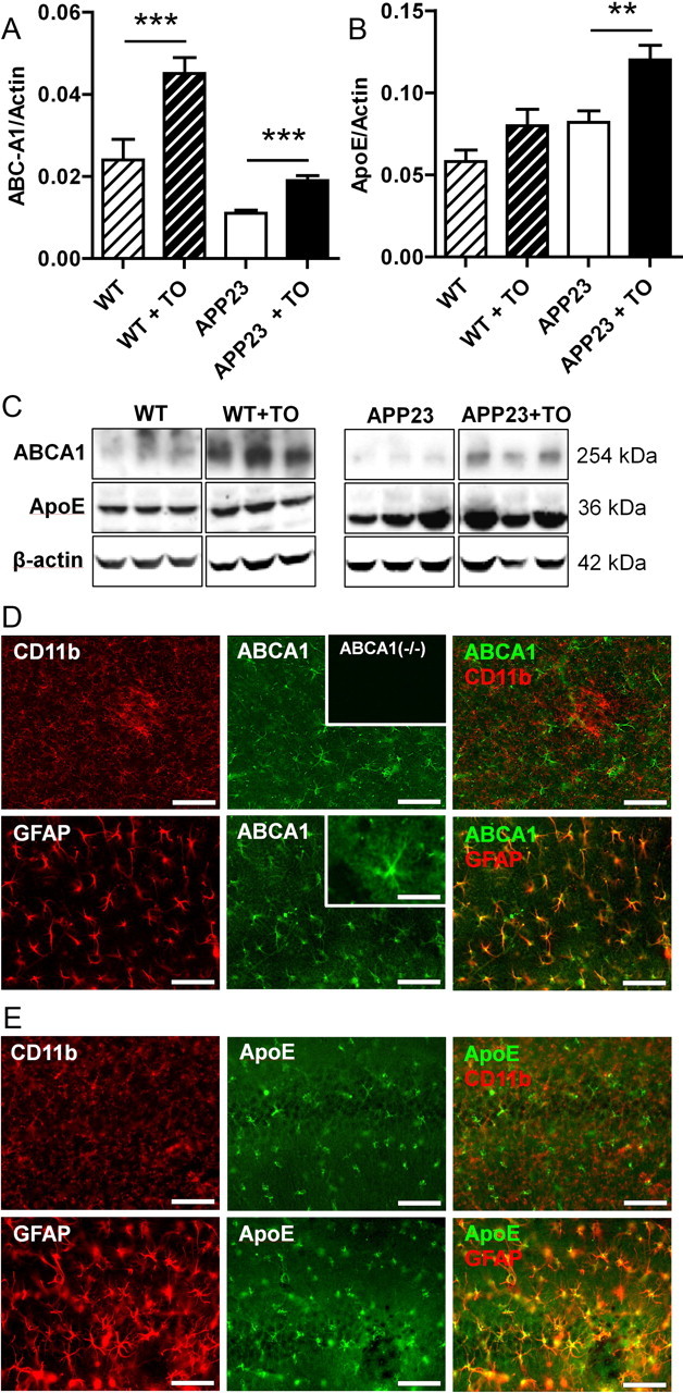
LXR activation positively regulates ApoE and ABCA1 expression in astrocytes. A, Densitometric quantification of Western blot detection of ApoE in brain lysates from APP23 and wild-type (WT) mice treated with either vehicle or TO901317 (TO). ApoE level was corrected for the level of the housekeeping protein actin. B, Densitometric analysis of Western blot detection of ABCA1 in brain lysates from APP23 and wild-type mice treated with either vehicle or TO901317. ABCA1 level was corrected for actin level. C, Representative images of ABCA1, ApoE, and β-actin Western blot detection in wild-type or APP23 mice treated with either vehicle or TO901317. D, E, Brain sections of vehicle-treated mice showing neocortex (D) or hippocampus (E) were immunostained for CD11b (microglial marker), GFAP (marker for astrocytic activation), and ApoE (D) or ABCA1 (E). The inset at the top of D shows absence of immunostaining in ABCA1 knock-out mice, while the inset at the bottom displays the ABCA1 immunostaining at higher magnification in the astrocytic arbors. Together, these immunostainings show that ApoE and ABCA1 expression is predominantly located in astrocytes but not in microglia in the murine brain. Asterisks indicate significant differences (ANOVA, Student's t test, n = 12, **p < 0.01, ***p < 0.001). Data represent means ± SEM. Scale bars, 100 μm, except inset, 25 μm.