
Figure 1.
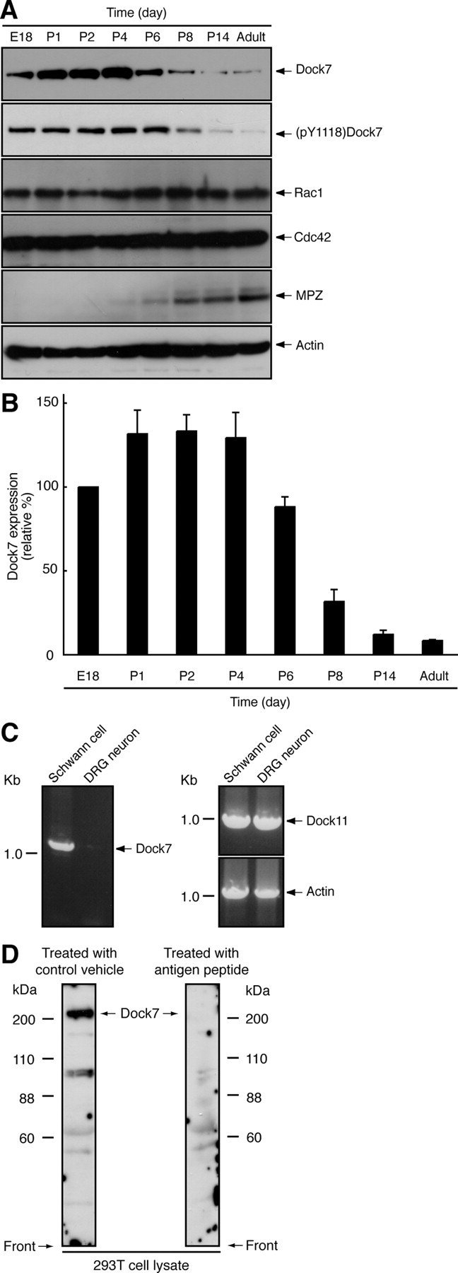
Dock7 is downregulated following sciatic nerve development. A, Tissue extracts were prepared from rat sciatic nerves on Embryonic Day 18 (E18) or Postnatal Day 1–14 (P1–P14), or upon adulthood, subjected to SDS-PAGE, transferred to PVDF membranes, and immunoblotted with an antibody against Dock7, (pY1118)Dock7, Rho GTPases Rac1/Cdc42, the myelin marker protein MPZ, or actin (as a control). B, The band intensities of an immunoblot with an anti-(pY1118)Dock7 antibody are shown as the relative intensities (n = 3). C, RT-PCR analysis shows that Dock7 mRNA is abundantly present in primary Schwann cells and very weakly present in primary DRG neuronal cells. RT-PCR analysis for β-actin and Dock11 was performed as the control. D, 293T cell lysates were immunoblotted with an antibody against a KLYPDGRVRPTRE peptide consensus in rat, mouse, and human Dock7 in the presence of an antigen peptide (right panel) or with an antibody in a control vehicle (left panel). In 293T cells, this antibody primarily recognizes >200 kDa of the full-length Dock7, which disappears in response to the treatment with antigen peptide. The lower bands (88–110 kDa) may correspond to the alternatively spliced variant of Dock7 (GenBank Acc. No. DQ309763) and/or may be degradation products of Dock7. Data were evaluated using one-way ANOVA (*p < 0.01).