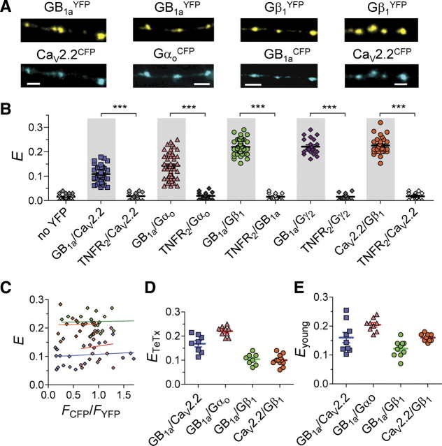
Figure 1.
GB1aRs, Gαoβ1γ2 G-protein subunits, and CaV2.2 channels are precoupled at single hippocampal boutons. A, Representative confocal images of pyramidal neuron axons in hippocampal cultures that were cotransfected with GB1aYFP/CaV2.2CFP, GB1aYFP/GαoCFP, GB1aCFP/Gβ1YFP and CaV2.2CFP/Gβ1YFP. Scale bars, 2 μm. B, FRET was detected between GB1aYFP/CaV2.2CFP (n = 33, N = 7), GB1aYFP/Gαo*94CFP (n = 53), GB1aCFP/Gβ1YFP (n = 45, N = 10), GB1aCFP/Gγ2YFP (n = 21, N = 4), CaV2.2CFP/Gβ1YFP (n = 31, N = 6) proteins under miniature synaptic activity at single hippocampal boutons. To verify FRET specificity, E was measured between the CFP-tagged proteins of interest and nonrelated TNFR2YFP. Error bars indicate SEM. ***p < 0.001. C, FRET efficiency is plotted for individual presynaptic boutons as function of CFP/YFP intensity ratio (FCFP/FYFP). No correlation was found: Spearman r is 0.14, 0.11, −0.08, and 0.05 for GB1aYFP/CaV2.2CFP, GB1aYFP/Gαo*94CFP, GB1aCFP/Gβ1YFP, and CaV2.2CFP/Gβ1YFP, respectively (p > 0.5). D, FRET was detected between GB1aYFP/CaV2.2CFP (n = 8, N = 3), GB1aYFP/GαoCFP (n = 9, N = 3), GB1aCFP/Gβ1YFP (n = 8, N = 3), and CaV2.2CFP/Gβ1YFP (n = 12, N = 4) proteins in nonreleasing TeTx-pretreated hippocampal boutons. E, FRET was detected between GB1aYFP/CaV2.2CFP (n = 9, N = 3), GB1aYFP/GαoCFP (n = 9, N = 3), GB1aCFP/Gβ1YFP (n = 10, N = 3), and CaV2.2CFP/Gβ1YFP (n = 9, N = 3) proteins in nonreleasing immature (4–5 DIV) hippocampal neurons.

