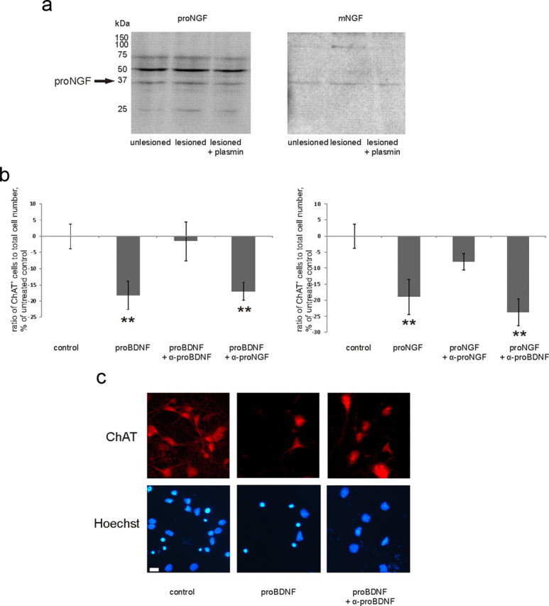
Figure 3.

a, Representative Western blot with antibody to proNGF (left), reblotted with the antibody to mature NGF (right). Band row at 35 kDa is recognized by both antibodies, which verifies it as proNGF. For quantification, see Figure 2f. b, To verify the function-blocking specificity of antibodies to proNGF and proBDNF, we applied proNGF and proBDNF in the absence or presence of antibodies to basal forebrain primary cultures (for details, see Materials and Methods). Each antibody specifically diminished the reduction in the number of ChAT-positive neurons induced by its cognate proneurotrophin, not having an effect on the response to another proneurotrophin. **p = from left to right: 0.027, 0.046, 0.015, 0.001, one-way ANOVA and Bonferroni's post hoc analyses; n = 5670 cells. Scale bar, 10 μm. c, Representative pictures from the experiment described in b. Error bars indicate SEM.
