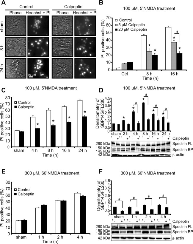
Figure 2.
Calpain inhibitor calpeptin protects neurons against excitotoxic apoptosis. A, Representative images of phase-bright and the merged image of PI- and Hoechst-stained neurons exposed to sham conditions or 100 μm NMDA for 5 min in the presence and absence of the calpain inhibitor calpeptin. Images were taken at selected time points (8 and 24 h after treatment). Scale bar, 10 μm. B, Dose–response assay. Neurons were treated with 100 μm NMDA for 5 min in the presence (5 or 20 μm) or absence of calpeptin for the indicated times. The number of PI-positive cells was expressed as a percentage of total cells in the field. Data represent means ± SEM from n = 3 separate cultures. *p ≤ 0.05 compared with NMDA-treated controls. #p ≤ 0.05 between NMDA-treated neurons in the presence of calpeptin (5 or 20 μm) (ANOVA, post hoc Tukey's test). C, E, Cortical neurons were treated with 100 μm NMDA for 5 min over 24 h (C) or 300 μm NMDA for 60 min over 4 h (E) in the presence or absence of calpeptin, and the extent of injury was assessed with Hoechst and PI. Data represent means ± SEM from n = 3 separate cultures. *p ≤ 0.05 compared with NMDA-treated controls (ANOVA, post hoc Tukey's test). D, F, Western blot and densitometric analysis comparing the levels of spectrin cleavage in neurons after NMDA treatments: 100 μm NMDA for 5 min over 24 h (D) or 300 μm NMDA for 60 min over 4 h (F) in the presence or the absence of calpeptin. β-Actin was used as loading control. Experiments were repeated three times with different preparations with similar results. Densitometric data are expressed as a ratio of the 145 kDa spectrin breakdown product (BP) and the 280 kDa full-length (FL) protein normalized to β-actin. *p ≤ 0.05 compared with NMDA-treated controls. #p ≤ 0.05 between NMDA-treated neurons in the presence or absence of calpeptin (ANOVA, post hoc Tukey's test).

