
Figure 5.
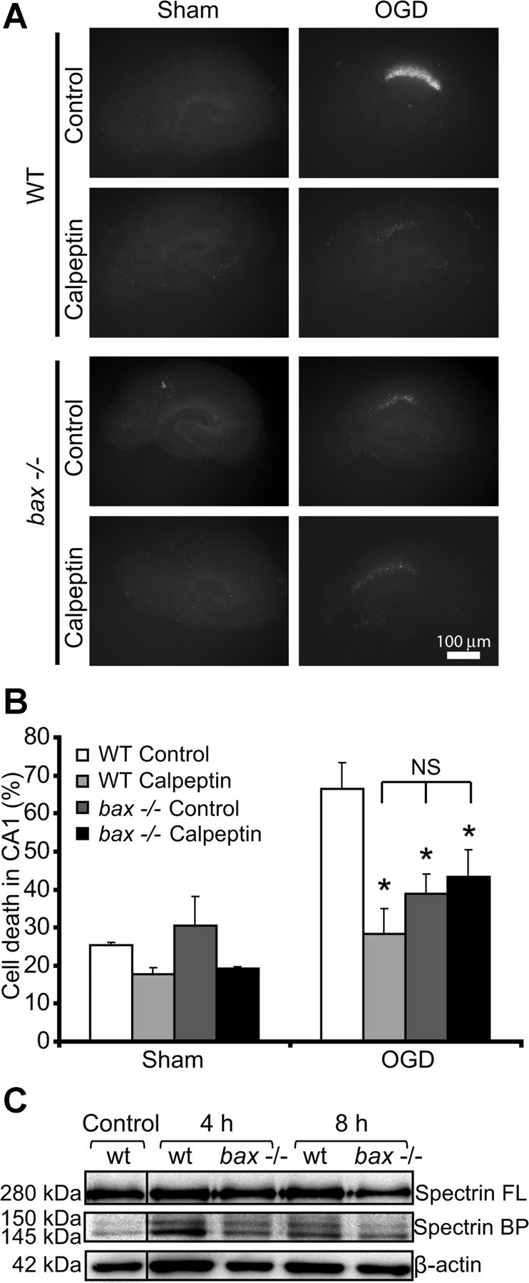
OHSCs isolated from bax-deficient mice show reduced calpain activation in response to OGD. A, Representative images of OHSCs derived from WT and bax−/− mice. The slices were sham exposed or subjected to OGD conditions for 45 min in the absence (control) or presence of calpeptin (20 μm) and allowed to recover for 24 h. Scale bar, 100 μm. B, Hippocampal slice cultures from WT and bax−/− mice were treated as described in A, and quantification of injury was assessed by PI staining 24 h after treatment (n = 5 slice cultures for each condition). Means ± SEM are shown. *p ≤ 0.05 compared with OGD-treated WT control (ANOVA, post hoc Tukey's test). C, Whole-cell extracts were prepared from WT and bax−/− hippocampal slice cultures treated to 45 min OGD and allowed to recover under normoxic conditions for the indicated time points. Spectrin cleavage was assessed by Western blotting. β-Actin served as loading control. BP, Breakdown product; FL, full-length.