FIG 2.
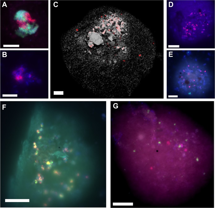
Epifluorescence micrographs of “Ca. Methanoliparia,” “Ca. Argoarchaeum” (GoM-Arc1), and “Ca. Syntrophoarchaeum” cells in oily sediment taken after CARD-FISH. (A to G) Visualizations of “Ca. Argoarchaeum” (GoM-Arc1) cells with the GOM-ARCI-660 probe (red) and bacteria (green) (A), “Ca. Syntrophoarchaeum” cells in consortia with the SYNA-666 probe (red) (B), “Ca. Methanoliparia” with the DC06-735 probe (red) in an immersed oil droplet (autofluorescence in gray) (C), “Ca. Methanoliparia” with the DC06-660 probe (red) in an immersed oil droplet (D), “Ca. Methanoliparia” cells (red) and bacteria (green; targeted with probe EUB388 I-III) (E), dual CARD-FISH staining of “Ca. Methanoliparia” cells with the specific probes DC06-735 (green) and DC06-660 (red) (F), and “Ca. Methanoliparia” cells (red, targeted with the DC06-735 probe) and ANME-1 cells (green) with the ANME-1-350 probe (G). (A, B, D, E, and F) Additional DAPI staining appears in blue. (D to G) Contours of oil droplets are visible by autofluorescence. Scale bars in all images, 10 μm.

