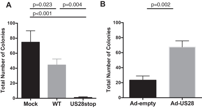
FIG 9.
US28 induces myeloid colony formation in CD34+ HPCs. (A) CD34+ HPCs were mock infected or infected with HCMV TB40E-GFP-WT or TB40E-GFP-ΔUS28 for 2 days. FACS-isolated viable GFP+ CD34+ HPCs were plated in Methocult H4434 at 500 cells/well and counted at 7 days. Data shown are average numbers of myeloid colonies per well for triplicate wells. Data are representative of three independent experiments. (B) CD34+ HPCs were infected with Ad-US28 or Ad-Empty (control). At 24 hpi, cells were plated in Methocult H4434 for 7 days. Data are representative of two independent experiments. Error bars represent standard deviations between three replicate wells per experiment. P values were determined one-way ANOVA (A) or by t test (B) and are listed as exact values. Replicate experiments are shown in Fig. S2.

