Figure 3.

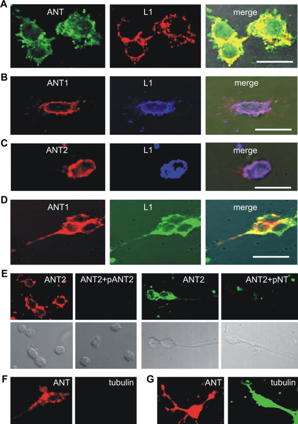
ANT1 and ANT2 colocalize with L1 at the cell surface of live cerebellar neurons. A–E, Live cerebellar neurons were stained with rat monoclonal L1 antibody 555 (A–D) and goat pan-ANT antibody (A), ANT1-specific (B, D) and ANT2-specific (C) antibodies, and ANT2-specific antibody that had been preincubated with peptides containing the N-terminal amino acids 2–13 of ANT2 (pANT2) or 1–28 of ANT1 (pNT) (E). F, G, Live neurons were stained with goat pan-ANT and tubulin antibodies (F), and fixed and permeabilized neurons were stained either with goat pan-ANT or tubulin antibodies (G). Confocal microscopy shows colocalization of ANT with L1 in the merged images (A–D). Scale bars: 10 μm.
