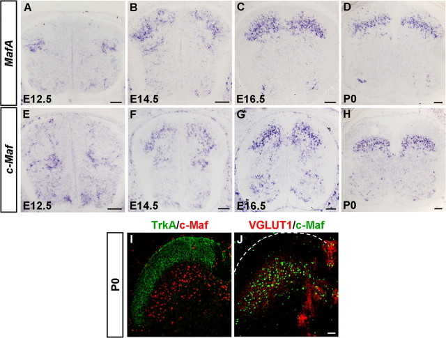
Figure 1.
Expression of MafA and c-Maf in the developing mouse spinal cord. A–H, In situ hybridization was performed on transverse sections of the spinal cord at various developmental stages using MafA and c-Maf genes as the probes. MafA- and c-Maf-expressing cells first emerged at E12.5 in the dorsal spinal cord (A, E). From E14.5 to P0, most MafA- and c-Maf-expressing cells were enriched in the laminae III/IV (B–D, F–H). I, J, Double immunostaining of c-Maf with TrkA (I) and VGLUT1 (J) was performed on sections of P0 spinal cord. c-Maf+ cells were found enriched in laminae III/IV (mechanoreceptive input). Scale bars: A–H, 100 μm; I, J, 50 μm.

