Figure 10.
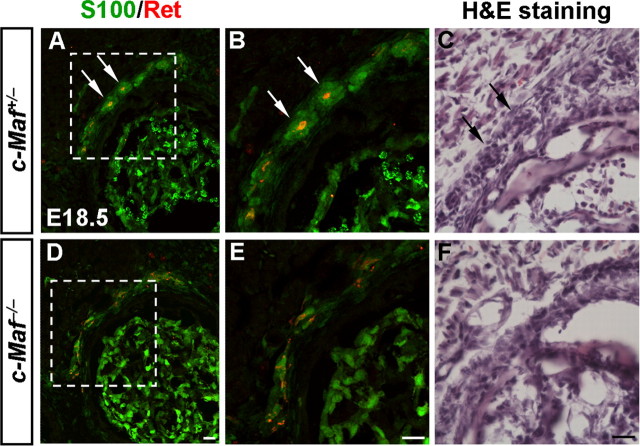
Defective development of Pacinian corpuscles in c-Maf deletion mice. A, B, D, E, Pacinian corpuscles (arrows) in the periosteum of the fibula of control (A, B) and c-Maf−/− (D, E) mice at E18.5 were visualized by double staining of S100 and Ret. Note the underdevelopment of Pacinian corpuscles in the mutant mice. B and E are higher magnification of the boxed areas in A and D, respectively. H&E staining of Pacinian corpuscles was performed in E18.5 control (C, arrows) and c-Maf−/− (F) mice. H&E staining is used here to rule out the potential confounding issue of decreased S100 and Ret expression in c-Maf deletion mice. Scale bar, 20 μm.

