
Figure 7.
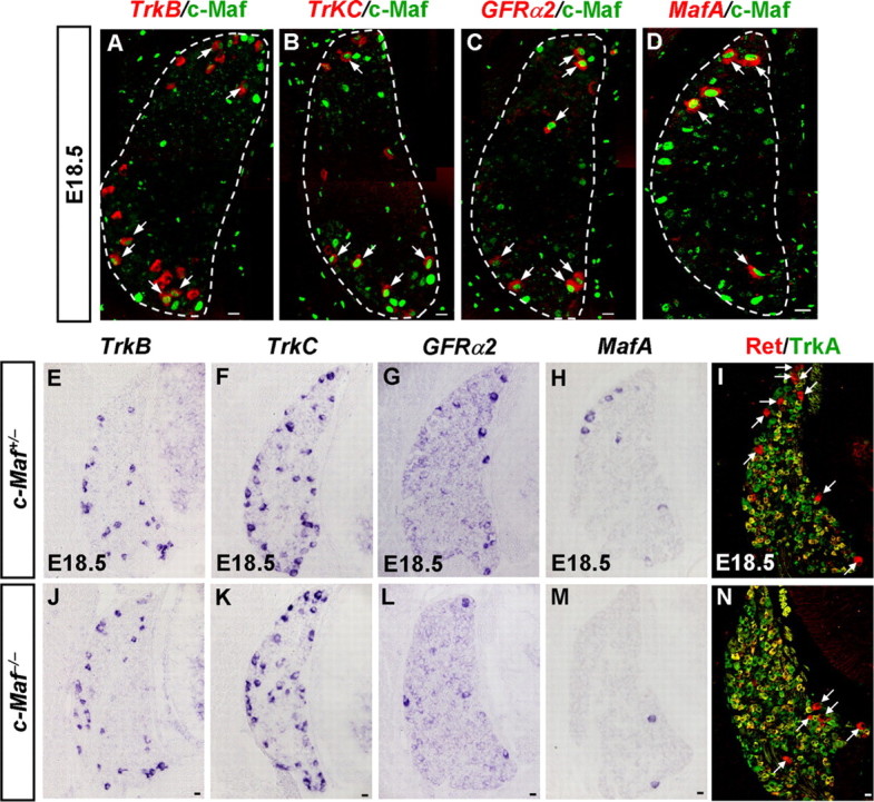
Defective differentiation of MafA+/Ret+/GFRα+ LTMs in c-Maf deletion mice. A–D, Coexpression studies of c-Maf with DRG neuronal markers at E18.5. Pseudo-color double staining of nuclear c-Maf protein (A–D, green) with TrkB mRNA (A, red), TrkC mRNA (B, red), GFRα2 mRNA (C, red), or MafA mRNA (D, red) was performed on DRG at E18.5. c-Maf+ neurons partially coexpressed TrkB or TrkC (A, B, arrows). Note that the majority of GFRα2+ and MafA+ neurons coexpressed c-Maf (C, D, arrows). E–N, In situ hybridization was performed on sections of DRGs from c-Maf+/− and c-Maf−/− mice at E18.5 with TrkB (E, J), TrkC (F, K), GFRα2 (G, L), and MafA (H, M) as the probes. Double staining of Ret (I and N, red) and TrkA (I and N, green) was performed on sections of DRGs from c-Maf+/− and c-Maf−/− mice at E18.5. Examples of large-diameter Ret+/TrkA− neurons are indicated by white arrows. Note that significant reductions of ∼55%, 70%, and 50%, respectively, in the numbers of GFRα2+, MafA+, and Ret+/TrkA− neurons were observed in c-Maf−/− mice. p < 0.001. Scale bar, 20 μm.