
Figure 1.
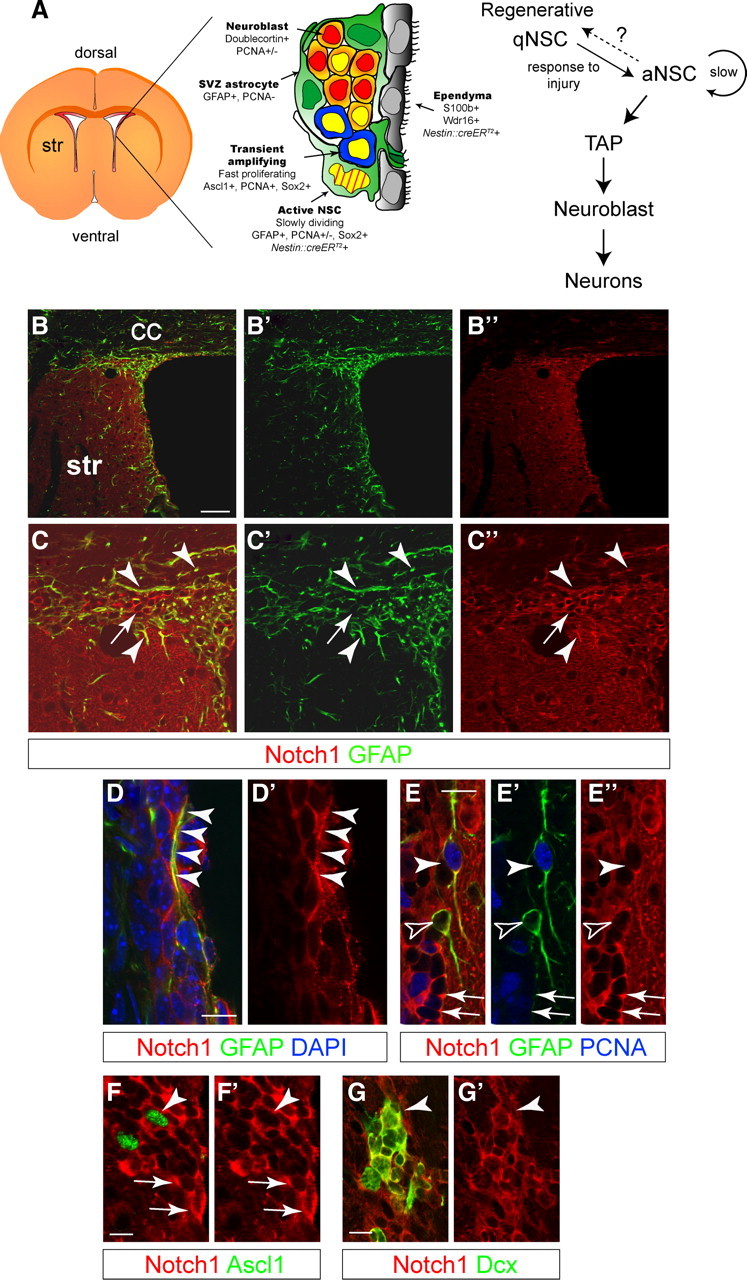
Notch1 is expressed in the adult subventricular zone niche. A, Schematic view of the SVZ and cell-type specific marker expression (adapted from Doetsch et al., 1999b). Hierarchical organization of the cells involved in adult neurogenesis in the SVZ. aNSC divide infrequently and generate TAPs, which in turn generate neuroblasts. Quiescent regenerative NSCs respond to signals (including injury), enter the cell cycle, and generate neurogenic aNSCs. It is unclear whether aNSC and regenerative qNSCs are independent states of the same cell and whether NSCs can shuttle between the two. Striped yellow nucleus is slow dividing, BrdU-retaining NSC; yellow nucleus is mitotically active cell. B, Low-magnification, confocal optical sections showing overview images of the lateral wall of the forebrain ventricle, including the SVZ. C, Notch1 protein codistributes with GFAP (arrowheads) but is also expressed by GFAP− cells (arrow). Higher-magnification images of sections in B. D, GFAP+ SVZ astrocytes express Notch1, including putative NSCs, with processes contacting the lateral ventricle (arrowheads) and interdigitated into the ependymal lining. E, Some astrocytes (GFAP+) are in the cell cycle (PCNA+, arrowheads) and some are not (GFAP+PCNA−, open arrowheads). SVZ astrocytes have lower levels of Notch1 compared with clustered putative neuroblasts (arrows). F, Neurogenic TAPs (Ascl1+) express Notch1 (arrowhead) comparable to other SVZ cells (arrows). G, Dcx+ neuroblasts (arrowhead) also express Notch1 (Nyfeler et al., 2005). Str, striatum; CC, corpus callosum. Scale bars: B, 100 μm; D–G, 20 μm.