
Figure 6.
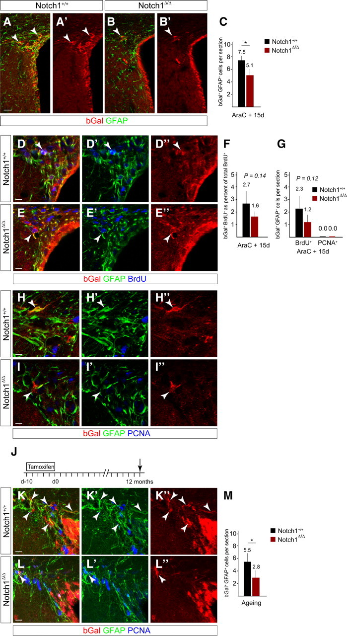
Notch1 ablation results in a reduction in GFAP+ dormant NSCs in the regenerated SVZ and during aging. A, B, bGal+ cells (arrowheads) in the regenerated SVZ 15 d after stopping AraC infusion are reduced in the Notch1-ablated (Notch1Δ/Δ) compared with control (Notch1+/+) animals. C, Ablation of Notch1 (Notch1Δ/Δ; n = 5) before regeneration results in a reduction in bGal+GFAP+ cells compared with control (Notch1+/+; n = 5) animals. D, E, BrdU label-retaining cells are present in the regenerated SVZ of control (Notch1+/+) and Notch1 cKO (Notch1Δ/Δ) animals and some express GFAP (arrowheads). F, bGal+ BrdU-retaining cells are slightly reduced in the regenerated SVZ following deletion of Notch1 (Notch1Δ/Δ; n = 5) compared with control (Notch1+/+; n = 5) animals. G, The number of bGal+GFAP+ BrdU-retaining cells is slightly reduced in the regenerated SVZ of Notch1 cKO (Notch1Δ/Δ; n = 5) compared with control (Notch1+/+; n = 5) animals. bGal+GFAP+ cells in the Notch1 cKO SVZ do not remain mitotically active (PCNA+) after regeneration. H, I, In both control and Notch1 cKO animals, GFAP+ cells return to a quiescent state (PCNA−) in the regenerated SVZ (arrowheads). J, 10 d TAM induction protocol in adult mice followed by 12-month chase (death, arrow). K–M, Ablation of Notch1 in young adult mice (Notch1Δ/Δ; n = 4) results in a reduction in bGal+GFAP+ cells when analyzed 1 year later compared with control mice (Notch1+/+; n = 4). Error bars are SD. Student's t test, *p < 0.05. Scale bars: A, 25 μm; D, E, H, I, K, L, 10 μm.