Figure 7.
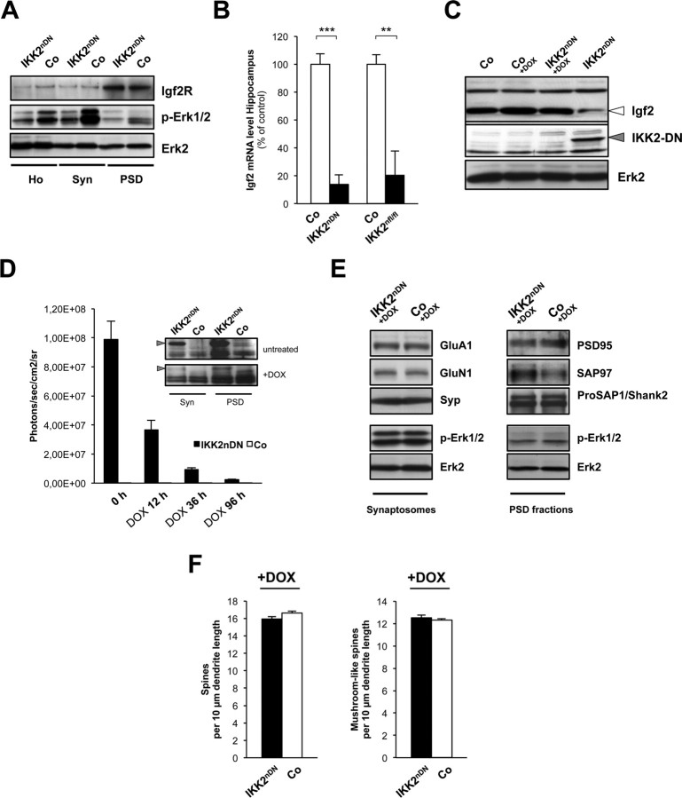
The IKK/NF-κB/Igf2 signaling system regulates synapse formation in vivo. A, Representative immunoblots of biochemical fractions from adult control and IKK2nDN mouse forebrain as indicated. Loading control, Erk2. B, Quantitative RT-PCR analysis of hippocampal Igf2 mRNA expression of control and IKK2nDN or control and IKK2nfl/fl animals. **p < 0.01; ***p < 0.001. C, Representative immunoblots of hippocampal Igf2 protein levels of control and IKK2nDN animals left untreated or treated with DOX. IKK2-DN indicates transgene expression, loading control: Erk2. D, Quantification of in vivo luciferase activity (in photons/second/centimeter2/steradian) in control and IKK2nDN brains before (0 h), 12, 36, and 96 h after DOX application. Inset, Immunoblots of synaptic fractions from control and IKK2nDN forebrains after 1 week of DOX application probed with anti-IKK1/2 antibodies. The arrowheads mark IKK2-DN transgene levels. E, Representative immunoblots of synaptosomal (left panel) and PSD fractions (right panel) from control and IKK2nDN forebrains 7 d after DOX application. Samples from 10 pooled forebrains are shown per lane. F, Quantification of spine density from secondary dendrites of CA1 hippocampal neurons from IKK2nDN and control mice. n = 5–10 cells from six independent male littermate pairs (left panel). Shown is quantification of mushroom-like spine density. n = 5–10 cells from six independent male littermate pairs (right panel). A–F, Co, Control; Ho, homogenate; Syn, synaptosomes; PSD, postsynaptic density fraction.

