Figure 7.
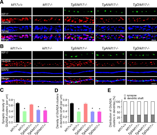
Intracellular localization of GluN2A. A, B, Primary cultures of hippocampal neurons were double-stained with anti-GluN2A/synaptophysin (A) or anti-GluN2A/MAP2 (B) antibodies. GFP-fused KIF17 (green) was present in TgS/kif17−/−, TgA/kif17−/− and TgD/kif17−/− neurons. Scale bar, 10 μm. C, Quantification of the synaptic density of GluN2A-positive clusters (colabeled with anti-synaptophysin) in dendrites. D, Quantification of the density of GluN2A clusters in dendritic shafts (colabeled with anti-MAP2). E, Comparison of the distribution of GluN2A clusters in dendritic shafts and synapses. Twenty neurons from three animals were examined for each genotype. Data are expressed as mean ± SEM (*p < 0.01; one-way ANOVA and post hoc test).

