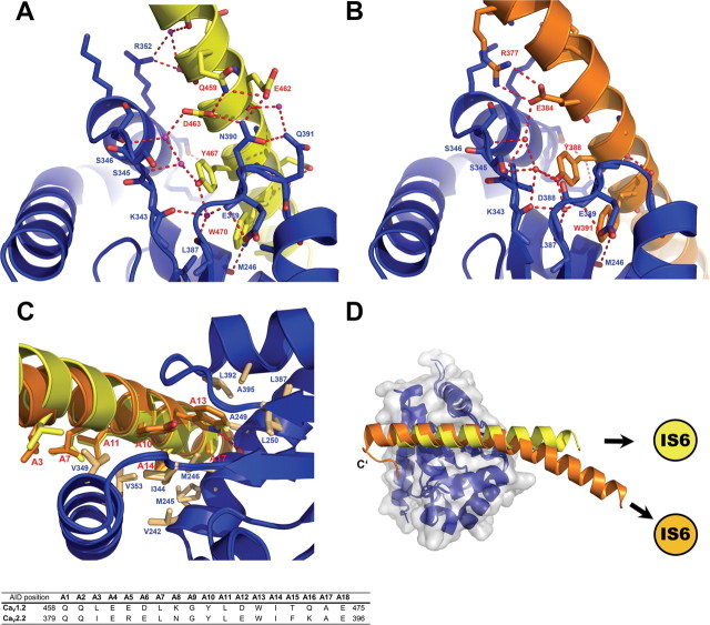
Figure 2.
The AID/CaVβ interface is structurally conserved between CaV1.2 and CaV2.2. A, B, Important polar interactions are conserved between the CaV1.2 (A, yellow) and CaV2.2 (B, orange) I–II linker/CaVβ interfaces. The GuK domain of CaVβ is blue. Important interface residues appear as sticks. C, Superposition of the AID/CaVβ interfaces of the CaV1.2 and CaV2.2 I–II linker/CaVβ2 crystal structures. Note the conserved position of many nonpolar residues (sticks) important for the interaction from both AID and Cavβ. D, Top view of the superposition of CaV1.2 and CaV2.2 I–II linker/CaVβ2 complexes.

