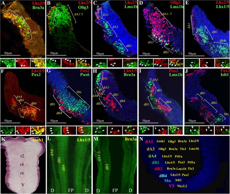
Figure 1.

The distribution of dorsal neuronal subpopulations in the hindbrain. A–J, Double-labeled immunofluorescence was performed on transverse-sections of E3 chick hindbrains at the level of rhombomere 4. The localization of each dA/dB neuronal subgroup is marked. The boxed areas are represented as enlargements in their different channels at the bottom of each panel. The arrows point at representative neurons in all channels. K–M, Flat-mounted views of E3 hindbrains following in situ hybridization (K) or immunofluorescence (L, M) staining. N, Summary of the distribution of the dorsal neuronal subtypes in the DV axis of r4. In all panels, antibodies and probes are indicated in their respective colors and scale bars are marked. D, dorsal; r, rhombomere; FP, floor plate; V, ventral.
