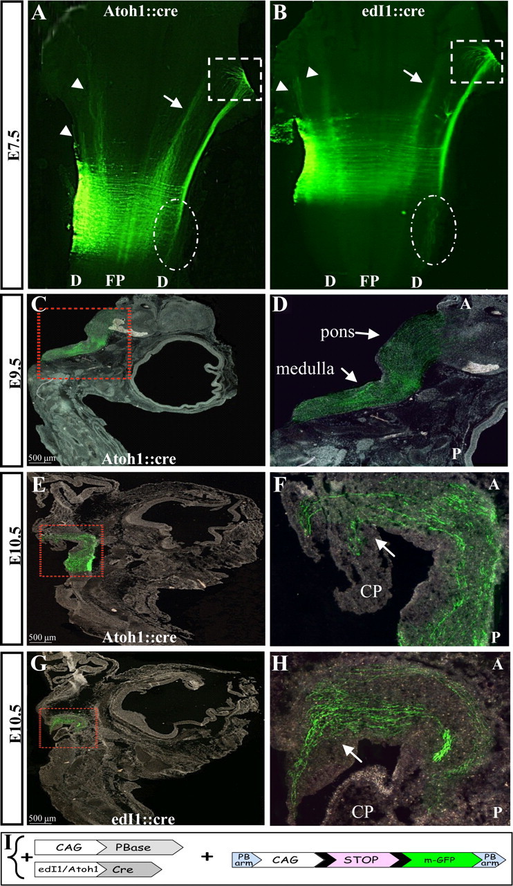
Figure 4.

Axonal projections of dA1 interneurons at E7.5–10.5. Flat mounted views (A, B) and sagittal sections (C–H) of embryos that were electroporated with the PiggyBac system to enable the incorporation of the conditional myristoylated GFP (mGFP) into dA1 cell genome. Axons were inspected at E7.5 (A, B), E9.5 (C, D) and E10.5 (E–H). A, B, arrows indicate the ascending contralateral medial tract, arrowheads indicate ipsilateral tracts, boxed areas represent the fasciculating axons of the ascending contralateral lateral tract and circled areas indicate descending contralateral axons. C–H, boxed areas in C, E, G represent higher-magnification views in D, F, H, respectively. Arrows indicate ascending axons in the medulla, pons, and cerebellar plate. In all images, stages and plasmids are indicated. A, anterior; P, posterior; D, dorsal; FP, floor plate; CP, cerebellar plate. I, A scheme of the constructs in which mGFP cassette is flanked by two PB arms (PB-LoxP-STOP-LoxP-mGFP-PB). The integration of the reporter cassette into the genome and the expression of mGFP in dA1 neurons is driven by CAG::PBase and Atoh1::Cre or edI1::Cre.
