
Figure 4.
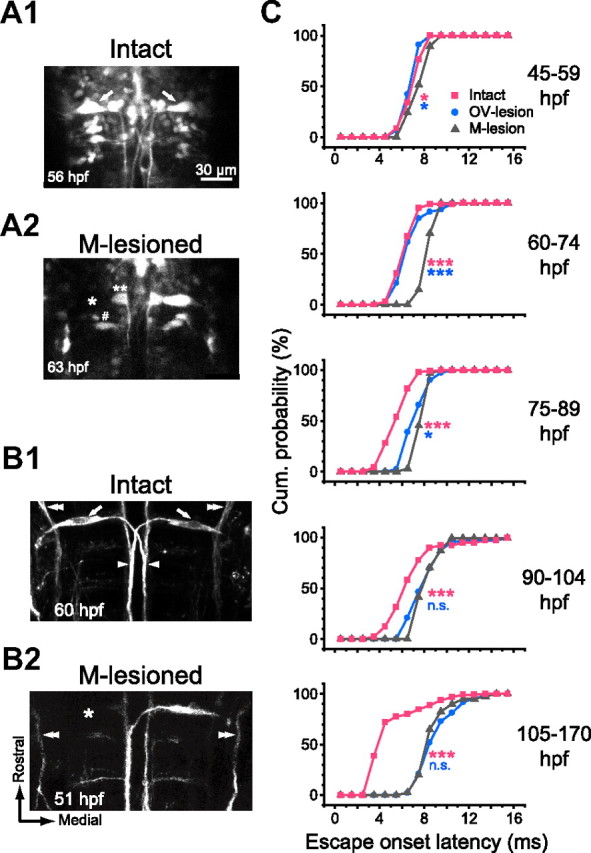
The M-cell is necessary to initiate fast escape with short latency. A, B, Selective M-cell ablation confirmed by GFP fluorescence (A) and immunostaining (B). A, GFP fluorescence images of hindbrain neurons including paired M-cells (arrows) in the Tol026 transgenic zebrafish (A1). The left M-cell was irradiated with laser pulses (A2). The M-cell body showed no recovery in fluorescence (asterisk), whereas the neighboring cells (number sign and double asterisks) still exhibited fluorescence, suggesting selective photo-ablation of the M-cell. B, Somata (arrows), lateral dendrites, and crossing axons (arrowheads) of both M-cells were labeled with the 3A10 antibody in an intact larva (B1). After laser irradiation of the left M-cell (B2), the 3A10 immunostaining of the left M-cell was not visible, whereas the descending trigeminal nerve (double arrowheads) remained intact. Scale bar in A is also applicable to B. C, Cumulative probability of onset latency of the tail flip response in M-lesioned fish (gray triangles) was superimposed on those for intact and OV-lesioned fish (pink squares and blue circles, respectively; reconstructed from Fig. 2, D and E). The latency was delayed significantly (pink asterisks) after ablation of the M-cell at all stages tested. Typically, responses with latencies <6 ms were never obtained after M-cell ablation. Note that the effect was even larger than that of OV lesion at 45–89 hpf (blue asterisks), suggesting that the M-cell is necessary to initiate escape with short latency in response to head-tactile stimulus. After 90 hpf, lesioning the OV or the M-cell results in a similar delay in onset latency (NS; p > 0.3), suggesting that the M-cell switches from being activated by head-tactile input to being activated by AV input in advanced larvae. *p < 0.05 and ***p < 0.001.