
Figure 6.
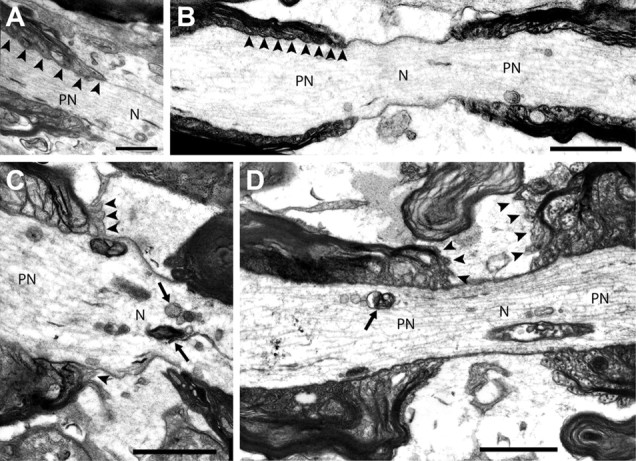
Dissociation of paranodal loops from the axolemma of myelinated neurons in the spinal cord following oligodendrocyte ablation. A–D, Electron microscopic assessment of nodes of Ranvier in longitudinal sections of the dorsal funiculus of the lumbar spinal cord revealed the nature of the interaction of paranodal loops and the axolemma of DT-challenged wild-type (A) and DT-challenged MBP-DTR mice (B–D). In all nodes examined in DT-challenged wild-type mice the paranodal loops closely abut the axon (A, arrowheads). A subset of nodes in DT-challenged MBP-DTR mice also maintained this regular structure (B); however, many nodes in these animals demonstrated alterations in axonal caliber, accumulation of organelles at the node and paranode (C, D, arrows), and unfurling or frank disconnection of paranodal loops from the axon (C, D, arrowheads). There was a 59.4% increase in the number of nodes described as abnormal (unpaired Student's t test). N, node; PN, paranode. Scale bar: (in D) A–D, 1 μm.