
Figure 5.
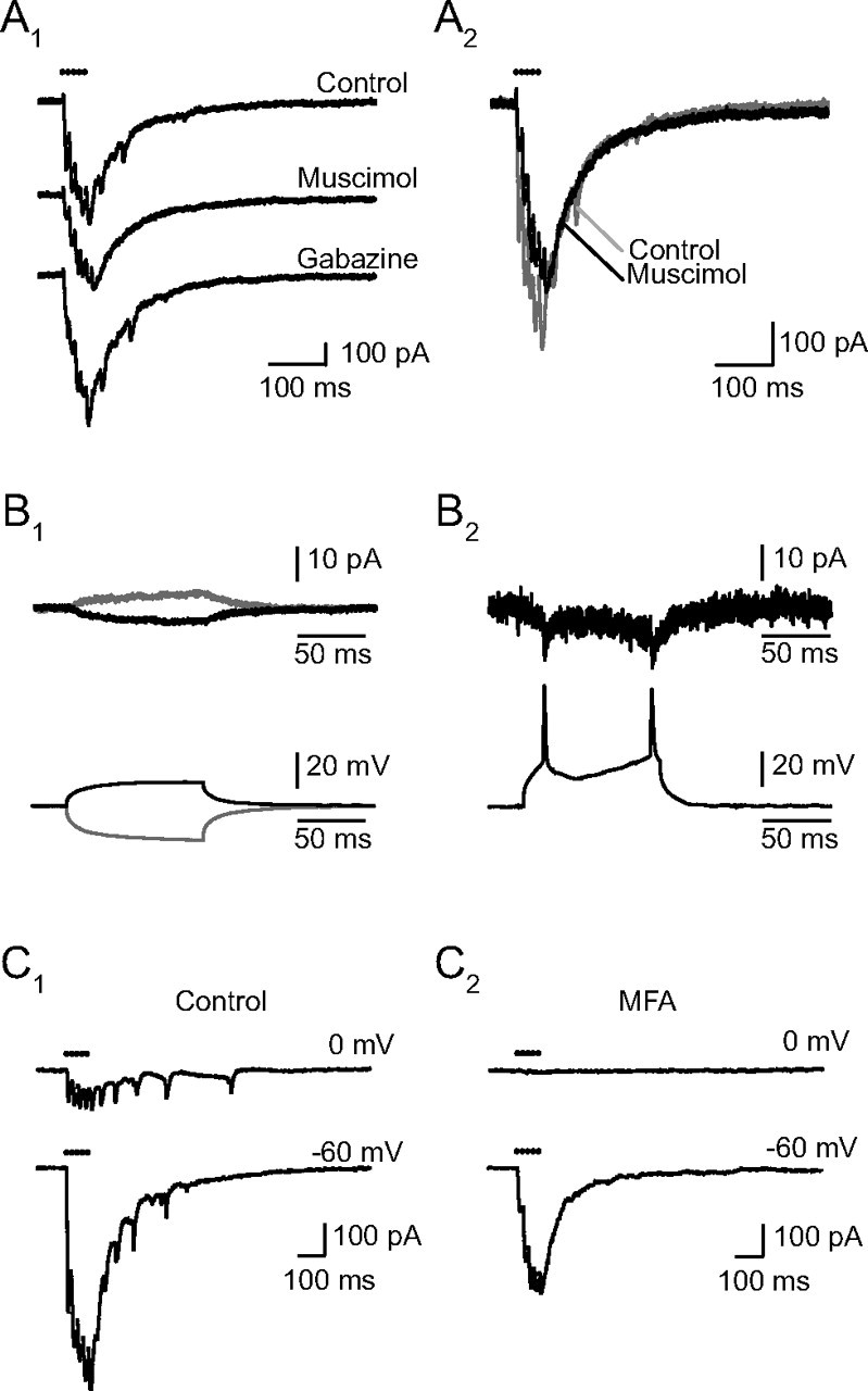
Electrical coupling between RCs. A, Muscimol eliminates the spikelets. A1, Muscimol (5 μm) suppressed both the initial and the late spikelets, reducing the peaks of the successive responses without altering the kinetics of the slow decay of the EPSCs. The nominal concentration of muscimol was 5 μm, but the records shown were taken just after the beginning of the perfusion, at which time the true concentration was certainly lower. Later the slow current also decreased as expected from a shunting of the coupled RC (data not shown). Washing muscimol and introducing gabazine (3 μm) led to the reappearance of the spikelets. A2, The superimposed traces before and after muscimol indicate that in its initial effect muscimol suppresses the spikelets but does not alter the decay of the eEPSC. B, Electrical coupling between RCs (identified by a response to the VR stimulation, data not shown). Two RCs were recorded simultaneously, one (Cs-RC) in voltage-clamp mode with a Cs-based internal solution containing QX314 (top recordings, Vhold = −60 mV) and the other (K-RC) in the current-clamp mode, with a K-based internal solution to allow the firing of action potentials (a DC current was injected to maintain the cell ∼−60 mV). B1, Pulses of current of various amplitudes were injected in the second RC and elicited an attenuated current in the first. In the two traces shown, 30 consecutive trials were averaged. The depolarizing pulse had an amplitude of 100 pA and induced a depolarization in the cell of +13.2 mV, the hyperpolarizing pulse's amplitude was −150 pA and induced a hyperpolarization of the cell of −19.2 mV. The depolarizing pulses induced an inward current in the first cell of −3.5 pA while the hyperpolarizing pulses induced an outward current of +4.4 pA. The electrical coupling can thus be estimated to 3.8 GΩ in the first trials, and to 4.4 GΩ in the case of hyperpolarizing pulses. B2, Supraliminar depolarizing current pulses injected in the K- RC and the corresponding spikelets recorded in the Cs-RC (single traces, no averaging). The spikelets are superimposed on the slow electrotonic response. C, MFA (50 μm) eliminates the spikelets and the inward current due to coupling (C1). In control conditions electrical coupling between the RC recorded from and neighboring RCs excited by the same presynaptic stimulation was revealed both by the presence of spikelets at −60 and at 0 mV and by the presence of a slow inward current at 0 mV. The spikelets recorded in this cell were exceptionally large. C2, After 75 min in MFA (50 μm), the spikelets had disappeared at both potentials. No synaptic current was observed at 0 mV. The response at −60 mV was reduced by half. The time constants of the two components of the decay were not changed, but the ratio of the fast over the slow component increased from 2.0 to 3.1. All records are single traces.