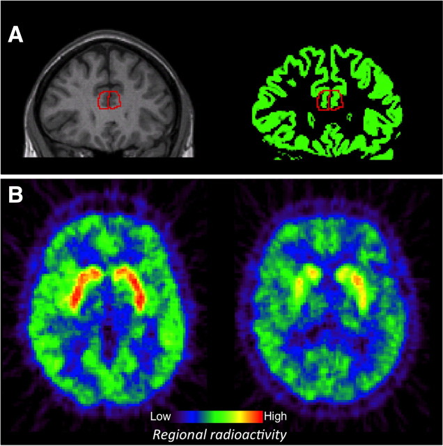
Figure 2.
Regions of interest for derivation of regional binding potentials were manually delineated on MRI images (A, left). Only segmented gray matter portions of the delineated regions were used in PET analyses (A, right). B gives an example of two PET images showing regional radioactivity in a young person on the left and an old person on the right.

