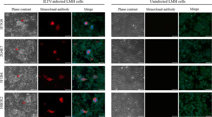
Fig 4. Recognition of gG by monoclonal antibodies in LMH cell cultures infected with the VFAR-043 strain.
Immunofluorescence assays were carried out using each monoclonal and anti β-Tubulin III antibodies. Nuclear staining was performed with DAPI mounting solution. Green and blue fluorescence correspond to β-Tubulin III and nuclear staining, respectively. Uninfected LMH cells were used as negative controls. Red arrows show cytopathic effect zones. Scale bars: 50 μm. Image magnification: 400x.

