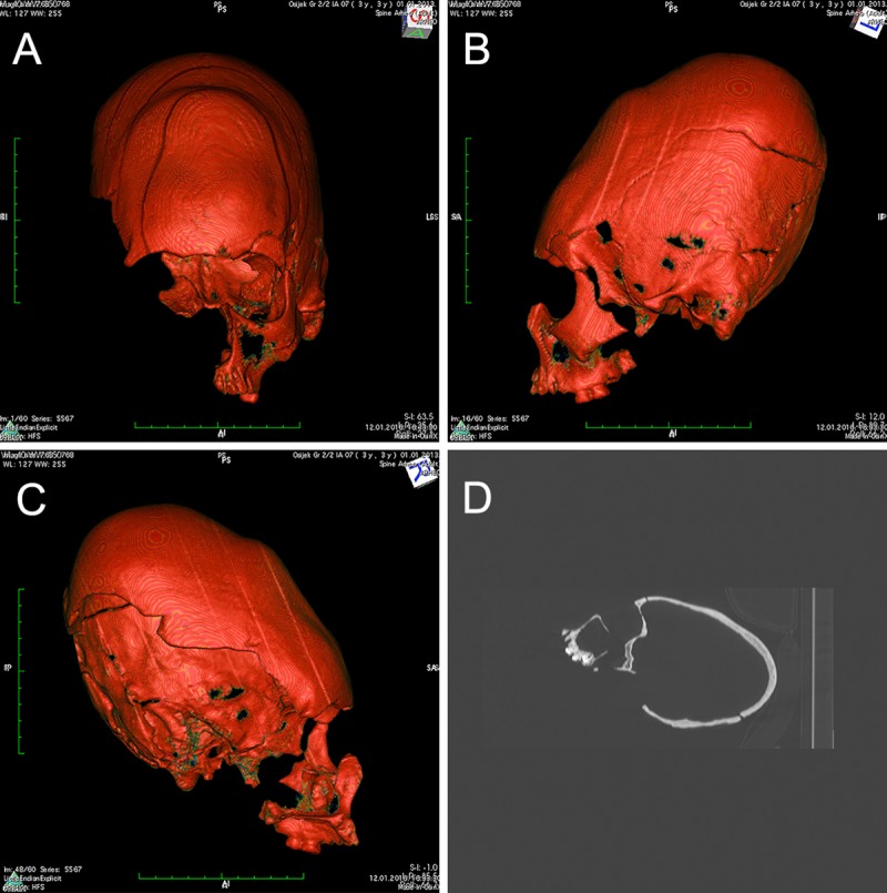
Fig 1.

A-C) CT reconstruction showing the artificially deformed cranium belonging to individual SU 259, frontal and lateral views. The shape axis is dislocated posteriorly above the Frankfort horizontal plane while the cranium exhibits a depressed and strongly inclined frontal bone strongly indicating tabular oblique type of deformation. D) X-ray of the same cranium (lateral view) showing a significant thickening of the posterior part.
