Fig. 5.
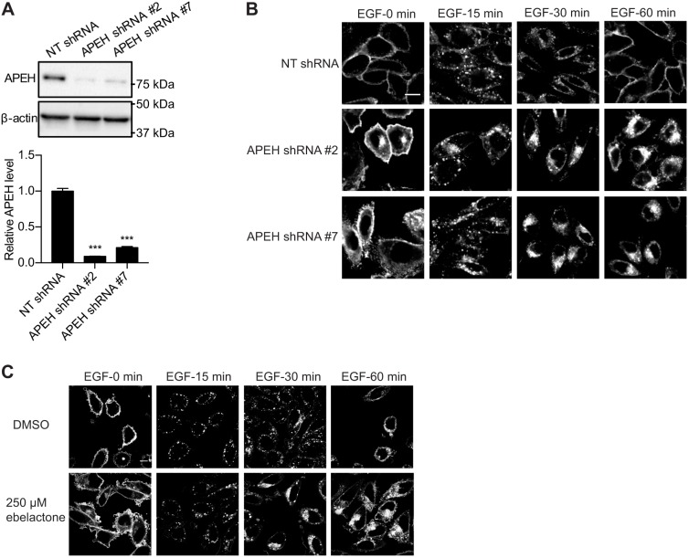
The endocytic recycling of EGFR is disrupted by APEH knockdown or inhibition. (A) CHO cells stably expressing mGFP–EGFR were transfected with NT shRNA or APEH shRNAs and lysed for quantitative immunoblotting. The level of APEH was normalized to the level of β-actin. Results are mean±s.e.m. (n=3). Representative western blots are shown. ***P<0.001 between APEH-knockdown and control APEH levels (one-way ANOVA). (B) Cells as in A were serum-starved for 2 h and incubated with 50 ng/ml EGF on ice for 20 min. Excess EGF was washed away and cells were incubated with fresh warm medium. Cells were fixed at different time points and imaged in a confocal microscope. Representative images are shown. (C) CHO cells stably expressing mGFP–EGFR were treated with vehicle (DMSO) or ebelactone. Cells were serum-starved for 2 h and incubated with 50 ng/ml EGF on ice for 20 min. Excess EGF was washed away and cells were incubated with fresh warm medium. Cells were fixed at different time points and imaged in a confocal microscope. Representative images are shown. Scale bars: 10 μm.

