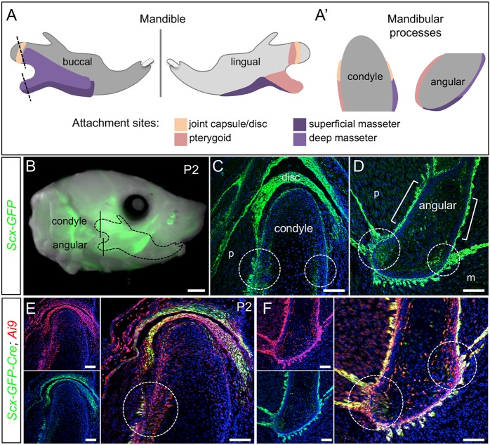
Fig. 1.
Scx-GFP and Scx-Cre mark tendon insertions in the mandible. (A,A′) Diagrams indicate attachment sites for the muscles of mastication and the temporomandibular joint capsule on the buccal and lingual sides of the mandible. Dotted lines indicate coronal plane of section for the condyle and angular process in A′. (B) Whole-mount of Scx-GFP at P2 identifies the craniofacial tendons (n=3). (C,D) Coronal sections through the condyle (C) and angular process (D) of Scx-GFP mice at P2, as indicated in B, show the insertion sites for force-transmitting tendons (circles) and muscle-anchoring tendons (brackets) (n=3). (E,F) Coronal sections through the condyle (E) and angular process (F) of Scx-Cre; Ai9 mice at P2 show the contribution of Scx+ cells to cartilage and bone at the tendon insertion (circles) (n=2). m, masseter; p, pterygoid. Scale bars: 1 mm (whole mount); 100 μm (sections).

