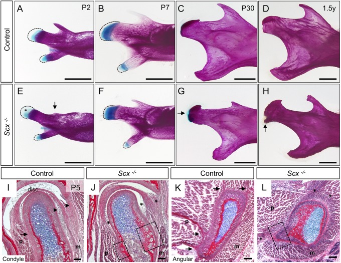
Fig. 2.
Scx regulates development of the mandibular processes and their tendon insertions. (A-H) Whole-mount skeletal preparations of mandibles from control (A-D) and Scx−/− littermates (E-H). Dashed lines mark the mesenchyme. (A,E) At P2, Scx−/− mandibles have an underdeveloped coronoid process (arrow) and ectopic Alcian Blue stain (asterisk) in the condylar mesenchyme (n=3). (B,F) At P7, the Scx−/− mandibles show precocious expansion of Alcian Blue stain in the condyle and angular process (n=2). (C,G) At P30, the Scx−/− mandibles exhibit ectopic Alcian Blue stain on the articular surface of the condyle (arrow), as well as a bulbous angular process (n=3). (D,H) By 1.5 years, the Scx−/− condyles develop a bony protrusion near the insertion site of the joint capsule (arrow) (n=2). (I-L) Coronal sections of the condyle (I,J) and angular process (K,L) stained with HBQ at P5. Tendon insertions in the controls (arrows) are disrupted in Scx−/− mice (asterisks). Articular disc attachments are disrupted in Scx−/− mice (arrowheads versus asterisks) (n=3 littermate pairs). Ectopic bone develops on the condyle and angular process at the sites of tendon insertions (boxes). m, masseter; p, pterygoid. Scale bars: 1 mm (skeletal preparations); 100 μm (sections).

