Fig. 4.
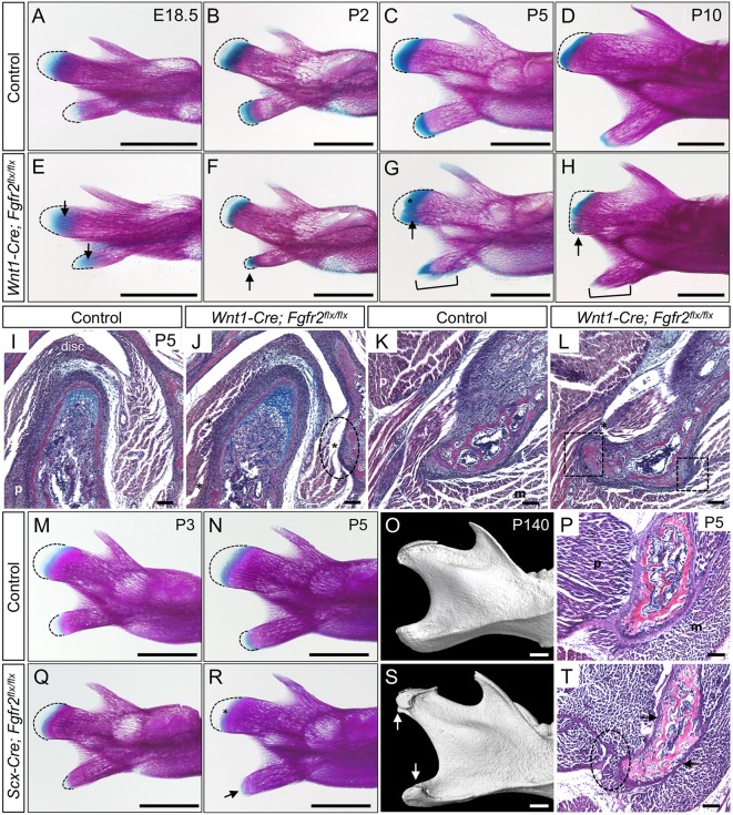
Fgfr2 regulates development of the mandibular processes and their tendon insertions. (A-H) Whole-mount skeletal preparations of mandibles from control (A-D) and Wnt1-Cre; Fgfr2flx/flx littermates (E-H). Dashed lines mark the prechondrogenic mesenchyme. (A,E) At E18.5, there is increased Alcian Blue in the mesenchyme of the Fgfr2 mutant condyle and angular process (arrows). (B,F) At P2, the mutant angular process is abnormally shaped and its mesenchyme is ectopically stained with Alcian Blue (arrow). (C,G) In the mutant at P5, ectopic bone spicules form at sites of tendon attachment (bracket) in angular process and ectopic Alcian Blue marks the growing front of the condyle (arrow, asterisk). (D,H) At P10, the mutant angular process is prematurely mineralized (bracket), and ectopic bone is seen at tendon attachment sites in the condyle (arrow). (I-L) Histological sections of the condyle (I,J) and angular process (K,L) stained with HBQ at P5. (I,J) In the mutant condyle, tendon insertion sites for the pterygoid, masseter and disc are incomplete (ellipse) compared with control. (K,L) In the mutant angular process, insertion sites for tendons (t) are incomplete (boxes) and contain ectopic bone (asterisk). (M-T) Whole-mount skeletal preparations at P3 (M,Q) show that the angular process of Scx-Cre; Fgfr2flx/flx mice is dysmorphic with premature loss of mesenchyme (outlined). At P5 (N,R), the cartilage of Scx-Cre; Fgfr2flx/flx mice is prematurely lost in the angular process (arrow) and condyle (asterisk). (O,S) μCT analyses at P140 identify ectopic bone on the condyle and angular process of Scx-Cre; Fgfr2flx/flx mice (arrows) (n=3 littermate pairs). (P,T) Histological sections of the angular process stained with HBQ at P5 show that tendon insertions are incomplete (ellipse) and the perichondrium is thin (arrows) in the mutant. n=3 littermate pairs for each. m, masseter; p, pterygoid. Scale bars: 1 mm (skeletal preparations); 100 µm (sections).

