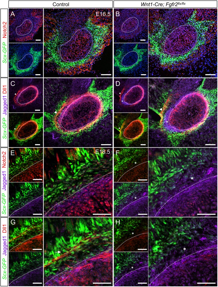
Fig. 9.
Loss of Fgfr2 alters expression of Notch pathway members at the tendon-bone interface. (A) In control at E16.5, Notch2 is high in the angular cartilage (dashed line), perichondrium and muscular connective tissue, whereas it is low in the tendon. (B) In Wnt1-Cre; Fgfr2flx/flx embryos, Notch2 is reduced throughout the angular process and nearby tissues. (C) Dll1 and Jag1 (Jagged1) overlap in the perichondrial region of the control. Dll1 is also localized to cellular extensions in the Scx-GFP+ tendon (arrowhead). (D) In the Wnt1-Cre; Fgfr2flx/flx angular process, Dll1 and Jag1 occupy distinct domains in the perichondrium (asterisk). The cellular extensions expressing Dll1 in the tendon were displaced from the perichondrial surface (arrowhead). (E) In the control angular process at E18.5, Notch2 and Jag1 overlap in the perichondrial region. Notch2 is lower in the developing tendon-bone attachment populated by Scx+ and Scx− cells. Notch2 is also detected in the muscular connective tissue near the myotendinous junction. (F) In the Wnt1-Cre; Fgfr2flx/flx angular process, Notch2 is reduced overall and Jag1 is expanded in the tendon-bone attachment, which is primary populated by Scx− cells (asterisk). (G) In the control angular process, Dll1 and Jag1 are co-expressed in the perichondrium. Dll1 is also expressed in cellular extensions in Scx+ cells of the tendon (arrow). (H) In the mutant angular process, Dll1 is reduced throughout (asterisk). Smaller panels show individual fluorescent channels with DAPI. n=3 littermate pairs for each. Scale bars: 100 µm in A-D; 50 µm in E-H.

