Figure 1.

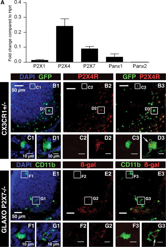
P2X1R, P2X4R, P2X7R, Panx1, and Panx2 expression in microglia. A, P2X1R, P2X4R, P2X7R, Panx1, and Panx2 expressions were analyzed on FACS-sorted SC microglia using qPCR. For each gene, the expression level corresponds to the x-fold change relative to the housekeeping gene Hprt. Note that P2X4R and P2X7R are the main P2XR transcripts expressed. The P2X1R transcript expression level is significantly (p < 0.01) lower than those of P2X4R and P2X7R. Note that low levels of Panx2 mRNAs were detected when compared with Panx1 mRNAs (≈13-fold significantly lower; p < 0.01). Error bars indicate SEM. B–D, P2X4R immunostaining in the ventral region of the SC of E13.5 CX3CR1eGFP mouse embryos. B1, Representative pictures of the SC ventral region. Note the accumulation of microglia (green) in the ventrolateral part of the SC. B2, P2X4R immunostaining. B3, Superimposed images shown in B1 and B2. C1, Enlarged image showing an example of eGFP microglia localized in the dorsomedial region of the ventral SC. C2, Note the lack of P2X4R immunostaining in the area shown in C1. C3, Superimposed images (C1 and C2) showing lack of P2X4R immunostaining within eGFP-positive microglia localized in the dorsomedial region. D1, Enlarged image showing an example of eGFP microglia localized in the ventrolateral region of the ventral SC. D2, P2X4R immunostaining in the area shown in D1 (red). D3, Superimposed images (D1 and D2) showing P2X4R immunostaining within eGFP-positive microglia localized in the ventrolateral region (arrow). Note that the staining is mainly located within the cytoplasm (single confocal section). E–G, β-Galactosidase immunostaining in the ventral region of the SC of E13.5 GlaxoSmithKline P2X7R−/− mouse embryos where the LacZ reporter gene has been inserted into the P2X7R gene. E1, Representative pictures of the ventral SC in E13.5 GlaxoSmithKline P2X7R−/− mouse embryos. Microglia were stained using CD11b antibody. E2, β-Galactosidase staining in the ventral SC. E3, Superimposed images shown in E1 and E2. F1, Enlarged image showing an example of microglia localized in the dorsomedial region of the ventral SC. F2, β-Galactosidase immunostaining in the area of the microglia shown in F1. Note the lack of galactosidase immunostaining. F3, Superimposed images shown in F1 and F2. G1, Enlarged image showing an example of microglia localized in the ventrolateral region of the ventral SC. G2, Galactosidase immunostaining in the area shown in G1. G3, Superimposed images (G1 and G2) showing galactosidase immunostaining within CD11b-positive microglia localized in the ventrolateral region (arrow; single confocal section), which indicates P2X7R gene expression in microglia. B1, C1, D1, E1, F1, G1, Cell nuclei are visualized with DAPI staining.
